The Art and Science of Cell Imaging
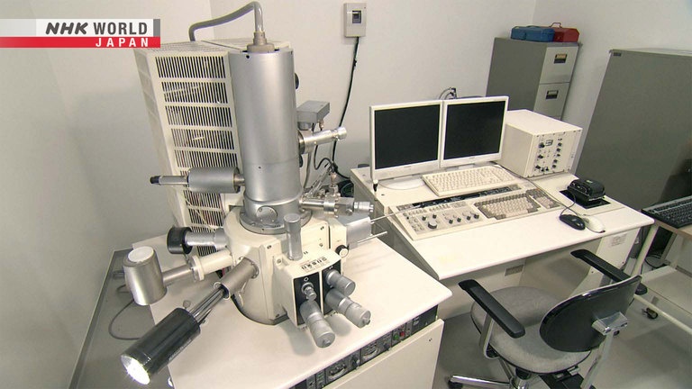
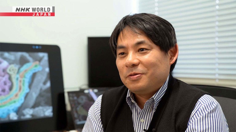
Have you ever looked at something up close? Really, really close? Micro-anatomist Daisuke Koga has looked even closer! He uses cutting-edge microscopes to visualize the insides of cells. As a leading expert in electron microscope images, his stunning high-tech photos have set new standards for both art and science. In this episode, he'll share his secrets for getting just the right picture. And we'll join him in trying out new equipment that creates full 3D images of the complex organelles that float inside cells. We'll also see how optical fiber lights can be used to ensure that nasal feeding tubes safely arrive in the stomach.